Endoscopic Ultrasound (EUS)
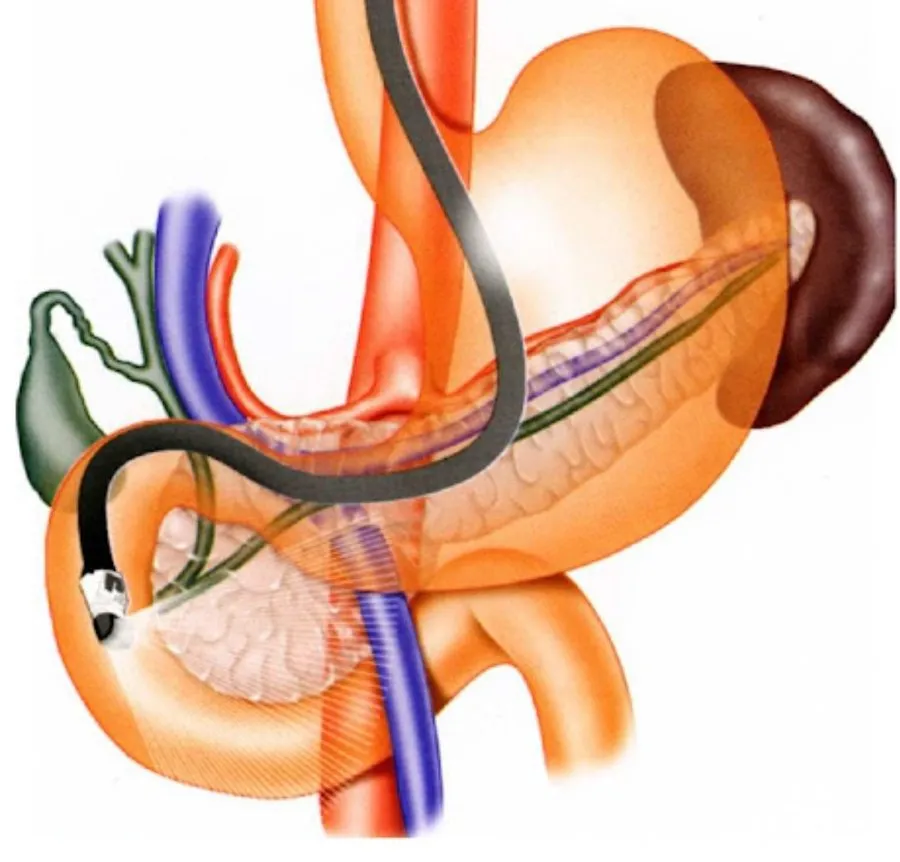
Digestive endoscopic ultrasound

Endoscopic ultrasound combines endoscopy (examining the inside of the digestive tract) with ultrasound (examining the wall and nearby organs using ultrasound waves).

What is endoscopic ultrasound?

Endoscopic ultrasound combines endoscopy (examining the inside of the digestive tract) with ultrasound (examining the wall and nearby organs using ultrasound waves). An ultrasound probe is brought close to the organ to be examined using an endoscope.

It is the most accurate examination of the walls of the esophagus, stomach or duodenum, and of nearby organs. It can diagnose stones, cysts and tumors of the bile ducts or pancreas. It also allows tissue that cannot be reached by other examinations (e.g. pancreatic tumors) to be sampled and studied under the microscope.

This examination is usually performed under general anesthesia. You must fast completely: no drinking for at least 3 hours before, and no eating or smoking for the 6 hours before the examination.

The examination uses a flexible instrument (echo-endoscope) inserted through the mouth or the anus. It is then positioned in front of the organ to be examined or sampled, depending on the indication. The examination lasts 15 to 20 minutes.

The instrument is cleaned and disinfected by an automated machine. Under validated cleaning, disinfection and sterilization conditions, no case of viral infection transmission (hepatitis B, hepatitis C, HIV/AIDS…) has ever been detected. You should be accompanied home after the examination and should not drive.

Apart from complications related to anesthesia, complications of endoscopic ultrasound are rare:

  • Diagnostic examination: complications are very rare, mainly perforation of the digestive wall
  • When samples are taken: very rare cases of infection, perforation or bleeding have been reported
  • When the pancreas is sampled: cases of acute pancreatitis have been observed